4D Scans Diagnose Heart Failure In 8 Mins

New MRI scans that use 4D images have been developed to be able to diagnose heart failure in just eight minutes.
Researchers from the University of East Anglia (UEA) have created technology that uses magnetic response imaging (MRI) to take 4D images of heart valves and blood flow. This has helped reduce diagnostic times from 20 minutes to just eight.
PhD student Hosamadin Assadi, from UEA’s Norwich Medical School, said: “This new technology is revolutionising how patients with heart disease are diagnosed.”
He explained the university teamed up with General Electric Healthcare to determine how reliable a new technique called Kat-ARC is as a diagnostic tool. It was found the scans not only reduce the amount of time it takes to get accurate pictures of the heart, but also measure the peak velocity of blood flow in the heart.
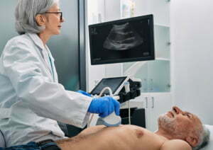
Lead researcher Dr Pankaj Garg, also from Norwich Medical School, stated the best way to diagnose heart failure is through an invasive assessment, or by ultrasound scan, which can be unreliable.
The 4D imaging, however, can look at the flow of blood in three directions over time, providing detailed information about the condition of the organ.
Dr Garg stated if the 4D MRIs become commonplace, “this will benefit hospitals and patients across the whole world”.
Causes of heart failure include high blood pressure, cardiomyopathy, a heart attack, congenital heart conditions, heart valve disease, and abnormal heart rhythms.
According to the NHS, heart failure impacts 900,000 people in the UK, with 60,000 new cases emerging every year.
For medical imaging systems, get in touch with us today.