A major new deep learning breakthrough that enables CT scans to segment tumours, lymph nodes and surrounding tissue has won an international prize.
The development, by postdoctoral researcher in medical radiation physics at Stockholm University Mehdi Astaraki, won his prize at the International Conference on Medical Image Computing and Computer Assisted Intervention (MICCAI).
Dr Astaraki’s work, which was focused on head and neck cancer, won the top prize in the Segmentation of Organs-at-Risk and Gross Tumor Volume of Nasopharyngeal Carcinoma for Radiotherapy Planning category, triumphing in a field of 395 entrants.
He presented his work at the event in the Canadian city of Vancouver in October, where his algorithms demonstrated outstanding performance, coming first in one assessment category and second in another.
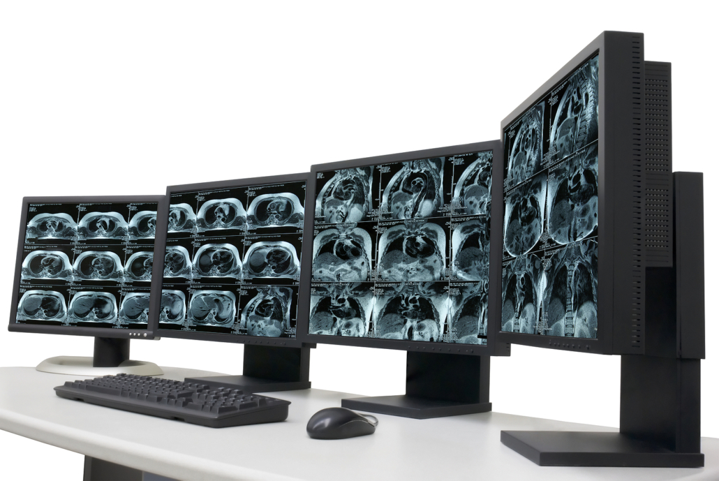
His work will mean medical practitioners sharing images using the technology will have better segmented details to study, meaning the data will help inform the treatment paths of patients more accurately and appropriately.
Indeed, a priority for Dr Astarki and his team will be to integrate his developed model with PACS systems, enabling it to play a vital role in planning radiation treatment. His presentation highlighted his desire for collaboration to become recognised as a crucial element of the development of medical image analysis.
Having participated in the contest in October and subsequently picking up the prize, he will present the findings in a news and views session in the spring, offering an update on the development of the system.
The development of new PACS-compliant medical imaging systems may also feature heavily next year, when the 2024 edition of MICCAI takes place in the Moroccan city of Marrakesh.
According to the organisation, the event will be “nothing short of groundbreaking”, although this is both for the new science on show and the fact it is the first time the annual event will have been held in Africa.