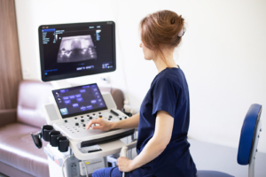
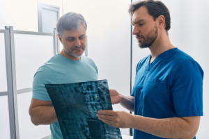
medical imaging doctor examining screen

As the use of medical imaging grows, the work involved can be more specialised and means that getting enough training to help make the best use of medical imagining solutions is a challenge.

However, new developments may help make this task easier, including a new innovation that follows on from one devised and launched only last year.

As Venturebeat reports, the launch last August of Stable Diffusion, a text-to-image foundational model, by Stability AI, prompted an idea from Stanford University radiologist Christian Bluethgen. He asked whether in fact it was possible to combine a genuine medical need with the creating of high quality images using basic text prompts.

The result was his collaboration with Stanford Graduate student Pierre Chambon, a mathematical and computational engineering researcher, which led to a study designed to establish the capacity of stable diffusion to generate X-rays. To their delight, they found it achieved this task very well.

All this, Mr Bluethgen, will help with the training of medics who might otherwise see very few scans relevant to their specialist area. He observed: “When you are working in a setting with scarce data, your performance correlates with experience – the more images you see, the better you become.”

Such a development may be just one indication of how better use of data technology is enabling medical imaging to be used with increasing effectiveness as a diagnostic tool, with improved training further boosting the capacity of specialists to identify medical problems when they emerge.

A further development in X-ray imaging may have emerged at the University of New South Wales in Sydney. In a paper published in the journal Nature Communications, researchers have revealed how a new algorithm has been devised that can enhance images of hydrogen fuel cells. It notes that the very same technique could be used to improve medical imaging.

world cloud medical

Cloud computing in healthcare could help balance the disparity between medical services around the world.

Over the last few years, use of cloud services has increased in the healthcare sector. They have advanced to store medical images and data, while artificial intelligence (AI) is being used more frequently to enhance treatments, diagnostic tools, and preventative procedures.

Dr Rowland Illing, director and chief medical officer, International Public Sector Health at Amazon Web Services (AWS), told ITP that cloud computing can improve access to technology. This makes it easier for healthcare providers with lower budgets to benefit from AI tools.

“The true potential of cloud computing in healthcare lies in its ability to democratise access to critical data, advanced tools for machine learning and artificial intelligence, and to make these resources accessible to researchers, developers, and pharmaceutical companies globally,” he stated.

As cloud computing does not require on-premises computer resources and large investments in advanced technology, it means it is more accessible for everyone.

According to the World Health Organization health equity is only achieved when everyone is able to “attain their full potential for health and wellbeing”.

It revealed nearly a third (30 per cent) of the global population are still unable to access essential health services. This was exacerbated by the Covid-19 pandemic, which caused disruption in health services in 92 per cent of nations.

The Universal Health Coverage (UHC) data stated that nearly two billion people currently face “catastrophic or impoverishing health spending”.


To find out more about cloud based medical imaging and how it can improve healthcare data storage in your team, give us a call today.

It will come as news to nobody that the NHS faces a tough winter. Fears of a ‘twindemic’ of high rates of Covid and flu infections, the backlog of operations caused by the pandemic remains and pay strikes among nurses and ambulance staff all speak of a health service under severe pressure.

Other outstanding cost concerns faced by the NHS include the high cost of agency staff, with doctors paid as much as £5,234 for a single shift according to data uncovered by the Labour Party in a Freedom of Information request, while many hospitals have outstanding maintenance issues as well. 

The question of just how much money could be needed to fix buildings, solve shortages of resources and settle pay disputes, alongside the question of where the money will come from, arises at a time when the UK is believed to have begun a protracted recession. 

Never has it been more important for cost-efficiency and the elimination of waste to be brought to bear on the NHS. That is where the low cost of cloud based medical imaging may be critical when keeping patient scans and other records, enabling easy access without high expense.

As Tech Radar reports, cloud storage specialists Backblaze have noted that hard disk drive prices have been falling, with prices now set to fall to as low as US$0.1 per GB of data. This compared to $0.11 in 2009.

Not only is this a rare example of something getting cheaper in a world beset by inflation, but also offers a considerable cost-saving opportunity to the NHS. While this alone will not solve all the problems, it will certainly help, not least by delivering better outcomes for patients, which is the whole point of the service in the first place.

While the NHS faces many challenges, it seems that at least the services the staff will use could include some cases of very low cost. 

3d ultrasound devices

A team at the University of Illinois has been given a $2m grant to develop devices that can add 3D medical imaging capability to traditionally 2D ultrasound systems.

The FASTER project, led by the Beckman Institute for Advanced Science and Technology, has been proposed as a way to make advanced three-dimensional imaging technologies more widely accessible without the need for dedicated and expensive CAT scanners or MRI machines.

Ultrasound, by contrast, is available in many diagnostic rooms along with X-rays, and thus is often one of the earliest forms of diagnostic imaging people will have taken when they see a doctor about a medical issue.

The concept behind ultrasound is similar to the system used by bats to perceive space and objects without sight. Bats emit a high-pitched ultrasonic sound wave that bounces off of objects and how that sound returns allows them to perceive and avoid obstacles in a process known as echolocation.

Ultrasound works similarly, typically using a probe or handheld devices to send a beam of ultrasonic waves around a part of the body, typically based on a known location such as a tumour or a foetus.

From this, the machine can determine the shape, size and location of the target in question and present that information. The only issue is that information is presented in two dimensions, which can make complex tissues, organs and tumours difficult to diagnose, often requiring several scans.

The proposed solution to this by the Beckman Institute is to use a clip-on device that attaches to the probe and instantly enables 3D ultrasound imaging in real-time.

A 3D ultrasound can capture the surrounding area, as well as the whole object, much in the same way a CAT scan does, and this can help doctors at a glance know exactly what kind of issue they are dealing with, in a way that is more cost-effective and thus more widely available.

Its first adaptation will be at the Mayo Clinic in Minnesota, with the hope that it can be more widely utilised based on its effectiveness.

Cloud Computing help

Disaster recovery is something every business or organisation needs to prepare for, whether it is a physical calamity on its premises like fire or flood, or a major data problem such as a power outage or cyber attack.

The capacity for cloud disaster recovery services to help is something many will have factored in when adopting the system. Buy storing and saving data in a particularly safe way it will ensure that medical imaging services are not disrupted and information from scans can still be sent to whichever practitioners need to see them.

Some may think an internet outage is an event that is unlikely to happen to them, especially with so many wireless services being available. But that is not always the case, especially in more remote corners of the UK.

Few places fit that description more than the Shetlands, which has just endured a loss of internet services as well as telecoms after two undersea cables connecting the islands to the mainland were damaged. These have now been fixed.

The incident followed another cable, linking the Shetlands with the Faroe Islands, also being damaged and affecting communications.

During this time, attempts to communicate with the mainland were futile, but any data stored in the cloud will have been safe, ensuring that once the cables were fixed it could be transmitted where necessary.

Because health services on the archipelago of 23,000 people are limited in scope, patients from the islands who need a scan – around 600 people a year – currently have to travel by ferry to Aberdeen to get one, so no scanning data would have been held up by the cable problems.

However, this situation is about to change. As Shetland News reports, NHS Shetland is moving its estates team to a site once occupied by the fish market in Lerwick to make space for the town’s Gilbert Bain Hospital to have a long-awaited MRI scanner fitted.

A lack of medical scans could have contributed to the exceptionally high level of excess mortality revealed in new official figures.

In 2022 over 650,000 deaths occurred, nine per cent more than the last pre-pandemic year of 2019. While there were more excess deaths in 2020 and 2021, Covid is the obvious explanation for those years. It was insufficient on its own, however, to explain the 2022 figures. 

However, the pandemic may have had an indirect impact by reducing the number of medical appointments, screenings and diagnoses in these years, preventing many people from getting the urgent, life-saving treatment they needed. 

While screenings may not have been cancelled, many patients will have been hesitant about coming forward for scans when infections were high and the NHS under severe strain.

The value of medical image sharing is that it enables the results of screenings to be easily accessed across different locations, which can have the benefit of enabling clinicians to provide the appropriate treatment for patients in a timely manner, often saving lives in the process. This is especially true for conditions like cancer.

An inability to access such images would be as bad as not having them in the first place, which can be the result of not having a scan to begin with – and it is apparent that the consequences of just such a scenario have played out with dire consequences for many patients over the past couple of years. 

Of course, there are other factors that may have contributed to what was one of the highest excess death rates in the last half century. The wider strains on the NHS may be a factor, not least at the end of the year when the worst flu season in a decade struck, itself a likely indirect consequence of reduced immunity after Covid restrictions prevented flu spreading much over previous winters.

The hot summer also contributed with the highest excess deaths during heatwaves among over-65s since comparable records began in 2004. However, this accounted for only 2,803 of the overall total, making it a minor factor.

heart ultrasound

In 2022, the biggest leap forward for medical imaging services was the adoption of artificial intelligence and machine learning to help doctors diagnose diseases in tissue samples and diagnostic scans,

This is why, for example, a system designed to detect different types of pastries is also used to scan for cancer.

However, a team from Rutgers University led by Partho Sengupta MD FACC and Naveena Yanamala PhD discovered a breakthrough in AI-assisted ultrasonic imaging that would enable an AI to detect changes in the function and structure of the heart at a molecular level.

This could be used at a very early stage to detect for signs of heart disease, potentially saving countless lives.

The system works by analysing patterns in echocardiogram imagery to determine at an expert level the types of conditions that lead to heart failure, establishing new biological markers that can help doctors plan appropriate cardiac treatment.

The biomarker can be applied to any current ultrasound device, which includes miniature hand-held ones that can fit in a pocket, and creates what the doctors call an “ultrasonic biopsy”.

There have been several similar stories of ultrasound being used in conjunction with AI to help detect heart disease, with one particularly fascinating study detecting weak heart pumps from data recorded by an Apple Watch, a consumer-grade smart device with limited health and fitness tracking.

With diseases like heart failure, lung disease, dementia and cancer, time is a critical factor in determining potential recovery. The longer the disease goes undiagnosed, the more of an effect it can have on people’s lives and the lower the chance of a full recovery.

Time is the difference between excising a benign tumour and being unable to operate due to cancerous tissue spreading to a vital organ.

This is a similar case with heart disease, as the sooner it is discovered, the more options are available to doctors.


man using 3d machine

Scientists have worked out how they can use artificial intelligence (AI) to help children in developing countries who have been born with cleft palates. 

Swiss researchers are using a 3D printer to create pre-surgical palate plates for babies in poorer countries, based on smartphone images of their cleft lips and palates, Swiss Info revealed. 

Computer software automatically generates a digital model of the palate, which can then be printed out quickly and easily. 

Andreas Muller, head of the Cleft Lip and Palate Treatment Centre at the University Hospital of Basel, designed the digital process together with scientists at the Department of Computer Science at the federal technology institute ETH Zurich. 

The procedure is currently being tested in India and Poland, and it is hoped it will make treatment more accessible for children in low-income states. 

Muller said: “As health workers, we feel frustrated faces with the fact that such ‘luxuries’ are unavailable to many families in those countries with scarce healthcare resources, training and a low number of healthcare workers.”

In higher-income nations, a team consisting of a paediatric dentist, a surgeon and anaesthesiologist create a plaster mould of the baby’s palate after they are born. An individualised plastic plate is then handcrafted by a dental technician before a qualified orthodontist implants this into the baby’s mouth and adjusts it regularly over a course of a six to eight months. 

After this, the cleft will have narrowed enough to only require a single surgery to close it. 

This surgery costs up to $3,500 (£2,880) in India, which is unaffordable for millions of people living there. 

If a cleft palate is not repaired, it can cause a number of health issues, including being unable to feed, hearing problems, a higher risk of tooth decay, and unclear speech. 


For medical imaging storage, take a look here. 

man working in lab

The constantly-adapting nature of Covid-19 made it difficult for medical staff to provide an accurate prognosis for hospitalised patients. However, scientists have now developed an artificial intelligence (AI) device that can offer a framework for continuous monitoring. 

Feinstein Institutes for Medical Research (FIMR) published their study in Nature Communications, having looked at more than 35,000 Covid-19 patients over the last two and a half years. 

Dr Theo Zanos, associate professor at the FIMR and Institute of Bioelectronic Medicine, led the research, which had to include the different changes in variants, waves and outcomes of the virus. 

He said: “Covid-19 was one of the most dynamic diseases we’ve witnessed in modern history and information about how to care for patients was constantly evolving.”

Scientists had to collect enough data to create a tool that accounted for the rapid developments. The auto-updating clinical device has been designed to assist clinicians in making decisions for patient care. 

While previous models have failed by not being able to consider fluctuations in patients’ characteristics and outcomes, the new model updates automatically when it detects a change in performance. 

President and chief executive officer of the FIMR Kevin J Tracey added the study was able to analyse data, and use modern technology to develop the tool and provide insights into the virus. 

It works by focusing on five early patient data points – age, serum urea nitrogen, lactate, serum albumin and red cell distribution width. By doing this, it was able to remain accurate across four waves and different variants. 

“Dr Zanos’ strategy provides a new model to study Covid-19 as a guide to clinical decision-making and better outcomes,” Mr Tracey added. 

For more information about cloud PACS system in healthcare, call us today.

digital imaging

Northern Ireland’s health and social care services are set for a major boost with news that a new imaging software system is to be deployed there. 

The system, developed by Sectra, is called NIPACS+ and will enable health trusts in Ulster to access patient scans and images from all over the region, irrespective of location, Digital Health Net reports

By bringing all medical imaging storage in the NHS in Northern Ireland under one single system, it means that for the first time every hospital from Belfast to Ballymena, from Limavady to Lurgan can all see crucial patient data at any time, no matter where the patient lives or has previously been treated or scanned. 

Other benefits will be the enabling of flexible working as staff who are based at home will still be able to see images, while new diagnostic hubs and training facilities will be established with the support of the system.

Chief executive at Belfast Health and Social Care Trust Dr Cathy Jack said: “This programme signals a major step forward in medical diagnostics for Northern Ireland.”

She added: “Moving to one imaging system will mean that from a single point of access healthcare professionals can instantly see all the imaging they need to support patient care.”

Managing director for Sectra in the UK and Ireland Jane Rendall said: “Northern Ireland is an example for the world in integrated diagnostics,” describing the project as an “extremely exciting one for Sectra to support”.

The development in Northern Ireland is a further sign of the growth in the use of medical imaging around the world, an expansion made easier by the accessibility to it provided by cloud storage systems and software.

According to new research from Pharmiweb.com, the global medical imaging market is set for compound annual growth of approximately 5.9 per cent between now and 2031.